



作为21世纪的绿色工程材料,Mg及其合金已成为重要的先进轻合金材料。镁合金因其密度低、比强度和比刚度高、生物相容性好、可回收性强及氢存储容量高等优异特性,在交通运输、国防军工、3C电子、航空航天、生物医学及能源等领域受到广泛关注[1]。
镁合金的生产加工通常涉及铸造、热处理、焊接或增材等多个复杂制备工序[2,3]。其中,在铸坯的铸造凝固过程或者构件直接成型的增材阶段,常常涉及晶体形核与生长、溶质分布、界面迁移及金属间化合物的形成等复杂的微观组织动态演变,以及缩孔/疏松、夹杂和溶质偏析等缺陷的形成。此外,为进一步提升镁合金性能,在加工和应用过程中常引入热场、压力场、电场、电磁场等多种外加物理场,进一步增加了微观组织演化过程的复杂性[4,5]。因此,在制备加工过程中,对微观组织的动态演化行为进行原位动态实时分析,对于调控合金成分、优化工艺参数以及改善力学和腐蚀性能至关重要。同时,在后续服役阶段,诸多由镁合金制造的零部件往往服役于复杂的工况环境中,一旦发生失效将可能导致重大的安全事故。因此,研究镁合金在复杂服役条件下微观组织的演化行为与损伤机制,已成为当前亟待解决的重要科学问题。
同步辐射光源的发展为解决镁合金研究中的关键科学基础问题和工程应用问题提供了独特而强大的技术手段。同步辐射光具有宽光谱范围、高亮度、高通量、高准直性、高偏振性、准相干性和脉冲时间结构等一系列优异特性,在综合的时空分辨率方面特性显著,其时间分辨率远高于传统电子显微镜技术[8,9]。尤其是同步辐射X射线具备的高能量和强穿透能力,使其可穿透毫米甚至厘米量级的金属材料,成为研究镁合金等金属内部微观组织的首选方法。基于同步辐射光源发展的多种X射线表征技术,如同步辐射X射线成像和X射线衍射等,已广泛应用于实时捕捉材料微观结构的动态演化过程。其基本思路是通过在光束线站集成各类原位样品环境装置,并结合X射线成像、衍射、小角散射及光谱等联用分析手段,实现外场(如力、热、化学环境)作用下材料内部精细结构、成分与缺陷演变的动态响应在线表征。例如,X射线成像技术可用于解析镁合金的微观组织形貌,而X射线衍射则可精确测量晶体内部的晶格畸变和应变分布。这些原位实验方法的建立,为深入揭示镁合金在制备、变形和腐蚀等复杂过程中的微观组织演化机制、力学行为及损伤机理提供了重要方法[10]。
本文综述了国内外基于同步辐射光源的原位实验装置发展现状,系统总结了利用这些先进技术研究镁合金在制备、加工及使役过程中微观组织和结构演变的最新进展。重点阐述了镁合金在凝固过程中多尺度微观结构的动态演化行为,加载过程中合金的微观变形和损伤机理,及腐蚀介质环境下的镁合金微观腐蚀动力学机制,重点探讨了同步辐射多维、多尺度、原位动态分析关键技术在镁合金研究中的作用、难点和重点。最后,对同步辐射技术在镁合金研究领域的未来发展方向进行了展望。
1 同步辐射原位研究技术开发及装置建设
同步辐射光源的高亮度、高准直性和宽谱段等优异特性,为实时原位研究材料在热、力或电化学环境下的动态响应行为提供了理想的研究手段。为开展各种环境下的材料微结构演变机理研究,首先需开发各类环境装置,以施加可调控的热、力、电磁或者化学环境,本节重点介绍国内外研究学者为同步辐射原位研究开发的新技术与搭建的专用装置。
Mathiesen等[11]最早基于第三代同步辐射光源开展金属凝固的研究工作,开发了X射线专用的凝固装置,该装置的冷却速率可在16.7~150 ℃/mm范围内调节,首次实现了对低熔点Sn-Bi和Sn-Pb合金的胞晶和枝晶动态生长行为的二维原位观察。此后,众多学者利用高能X射线二维原位成像开展了金属原位凝固过程组织演化研究。其中,Abou-Khalil等[12]基于欧洲同步辐射光源,开发了Bridgman炉(装置结构如图1a和b[12]所示),可用于两种凝固方式的实验:一种是通过电机驱动坩埚以1~50 mm/s的速率下拉,炉内热梯度保持在15~30 ℃/cm;另一种是通过降低加热器温度实现凝固,降温速率可控制在0.5 ℃/min,该装置在室温下的极限真空度可达10-9 bar (10-4 Pa)。利用该装置该团队实现了Al-14%Sn (质量分数,下同)合金的定向凝固实验二维原位成像表征。近年来,随着同步辐射光源和原位实验技术的不断发展,基于同步辐射X射线三维成像技术已能够实现对高温金属熔体三维组织及其随时间演变的原位表征。例如,英国曼彻斯特大学研究团队[13]利用该技术在0.05 ℃/s的慢冷却速率下,原位研究了镍基、铁基和钴基合金中三维枝晶形态的演变过程。实验装置如图1c[13]所示,该装置最高温度可达1300 ℃,极限冷却速率可达10-2 ℃/s,控温精度为±0.5 ℃。
图1

图1
基于同步辐射X射线的原位凝固与力学加载装置[12,13,15,16]
Fig.1
In situ device for solidification and mechanical loading based on synchrotron radiation X-rays
(a, b) 3D drawing (a) and photo (b) of Bridgman furnace[12] (UHV—ultra-high voltage)
(c) schematic of the environmental solidification cell setup at the I12 beamline of Diamond Light Source[13]
(d) in situ ultra-high temperature tensile loading device[15]
(e, f) schematic of thegeometries of specimens (e) and in situ mechanical rig for synchrotron X-ray diffraction experiment at I12 beamline of Diamond Light Source (f)[16] (ETMT—electro-thermo-mechanical tester. L—load direction, ϕ—azimuth angle, 2θ—diffraction angle)
为实现材料内部力学损伤动态演化过程的原位观测与原位三维动态(四维)表征,需研发与光束线站高度兼容的专用微型原位加载装置。此类装置的设计受多项关键因素的制约:首先,主体结构需采用对X射线无遮挡的布局,以保证后续图像重构质量;其次,其尺寸与总质量须满足线站旋转平台的承重限制;此外,还需综合考虑线站X射线的最优能量、材料本身的吸收特性及其基本力学性能,以确定合适的加载形式与载荷范围。自20世纪90年代以来,随着全球主要同步辐射光源的快速发展,研究人员开始系统研制专用的原位加载实验装置。Buffière等[14]较早开展了同步辐射成像下的原位拉伸研究,研制出与欧洲同步辐射装置ESRF ID19线站兼容的原位拉压试验机。该设备的主要参数包括:拉伸速率5 × 10⁻⁴~1.0 mm/s,有效载荷50~5000 N,整机质量约6 kg,高度约30 cm。利用该装置,他们对常温下Al/SiCp复合材料拉伸载荷下的裂纹扩展与断裂行为进行了原位成像研究,成功获取了材料内部SiC颗粒断裂及其与基体脱黏的三维图像。在超高温环境下材料内部损伤演化的研究方面,Ritchie研究组[15]取得了突破性进展,设计了一台兼容于先进光子源的原位超高温拉压试验机(图1d[15]),借助该装置,研究人员系统研究了Cf/SiCm和SiCf/SiCm复合材料在常温和极端高温条件下的三维损伤行为与断裂机制,该装置温度可达1750 ℃,视场中心5 mm内的变化仅为150 ℃。这项研究首次实现在超高温与外加载荷条件下对材料内部组织和力学行为的实时动态三维表征。此外,欧洲同步辐射光源和英国曼彻斯特大学也开发了类似高温力学装置,但在力学装置的加载结构上略有不同。
为研究电磁场作用下不透明合金凝固过程中晶粒的形核与长大行为,揭示其晶体生长的动力学机制,大连理工大学王同敏团队开发出一系列可在不同环境下研究金属凝固的原位装置,实现试样高温在线加热(800~1500 ℃),并可独立调控温度梯度和生长速率(图2a~d[17,18]),实现了电磁场在线施加技术,成功解决了磁场与电流装置、真空凝固控制系统、成像样品及同步辐射X射线光路之间的兼容性问题,克服了热与电相互干扰的难题。在此基础上,构建了用于实时观测电磁场下合金凝固晶体生长行为的高速成像系统,并建立了相应的原位动态数据定量分析方法。此外,该团队联合上海交通大学和哈尔滨工业大学,共同研制了适用于金属材料多物理场耦合制备与高温定向凝固的实验装置,可在0.8 T强磁场或高温度梯度条件下,实现对金属合金(包括高温合金、铝合金等)材料的晶体形核与生长动态演化过程的原位观测。
图2

图2
电磁场作用下基于同步辐射光源的凝固行为实时成像装置示意图[17,18]和照片及多功能/多环境服役原位加载装置
Fig.2
Real-time imaging device for electro-magnetic solidification behavior based on synchrotron radiation source and multi-functional/multi-environment service in situ loading device
(a, b) schematic (a)[17,18] and on-site photo (b) of the solidification device integrating electromagnetic fields (CCD—charge coupled device, DC—direct current)
(c) in situ high temperature vacuum solidification furnace at Shanghai Synchrotron Radiation Facility (SSRF)
(d) in situ 4D high temperaturesolidification furnace at SSRF
(e) physical diagram of the in situ 4D loading device developed based on the imaging line station at SSRF
(f) schematic showing the structure of a mechanical rig for in situ microdiffraction experiment at SSRF
金属部件常服役于复杂动态与交变载荷、极端高/低温或温度交变、腐蚀环境以及多种因素耦合的恶劣工况中,开展(近)使役环境下材料的微结构演变机理对于零部件的安全服役具有重要意义。王同敏团队依托上海同步辐射光源(简称上海光源,SSRF)研制了适用于极端环境(高温、低温及腐蚀等)的样品环境装置。成功开发出的极端服役热-力耦合环境材料损伤原位成像评价装置(图2e),可在-198~1680 ℃宽温域内,对材料在拉伸、压缩或剪切等复杂热-力耦合加载作用下的变形和损伤机制开展研究。此外,该团队还联合西安交通大学、南京工业大学、哈尔滨工业大学和上海光源,共同研制了一套用于材料微结构与应力分析的同步辐射微衍射原位环境装置(图2f),能够实现高温(约1200 ℃)力学加载、低温(-120 ℃)力学加载以及应力-腐蚀介质等多种耦合环境下的原位衍射实验。该装置可提供力学加载、温度与腐蚀介质多场耦合的原位实验条件,并借助同步辐射白光Laue微衍射技术,对金属材料在多因素作用下的微观应力、晶体取向分布以及微观结构(如位错、晶界和第二相等)实现定量化与可视化分析。
2 镁合金凝固过程的组织演化机理研究
铸造、焊接等传统的加工工艺以及当前较为先进的增材制造均涉及复杂的凝固过程。通过将同步辐射X射线表征技术与原位装置相结合,能够直观呈现金属液/固相变过程中微观组织的动态演变过程,从而为探究组织的演变机理以及通过调控工艺参数优化金属性能提供宝贵数据。
金属在铸造凝固过程中,会形成多种微观组织(如枝晶、共晶组织以及第二相等),其中,枝晶是铸造镁合金中最主要的微观组织,其形貌演化主要源于相变过程中固/液界面的失稳现象。枝晶的微观结构及其取向对镁合金的性能具有显著影响。
枝晶的形貌和取向受多种因素影响,如温度梯度、冷却速率、溶质元素及异质颗粒等。利用同步辐射X射线透射成像技术可快速获取镁合金凝固过程中枝晶形貌的动态演化。国内外研究学者利用同步辐射X射线二维/三维/四维成像技术从多个维度对枝晶生长的形貌和生长方向进行了深入解析。Casari等[22]对Mg-Nd-Gd-Zn-Zr合金在不同冷却速率和合金元素条件下的枝晶形貌变化进行了二维动态成像原位表征,发现在接近等温凝固条件下,商用Mg-Nd-Gd-Zn-Zr合金中的初生α-Mg等轴枝晶会经历形态转变,当冷却速率低于0.075 K/s时,生长模式从三维各向同性突然转变为沿<11
基于三维表征的分析虽从实验上验证了基于凝固理论所提出的枝晶生长模型,但仍存在一定的局限,因为组织的形成包含了复杂的动力学生长与演变过程。同步辐射四维(三维+时间)成像技术的发展为捕捉合金凝固过程的微观动态演变提供了重要分析方法。王同敏团队[28,29]构建了活泼镁合金的同步辐射原位四维成像凝固关键技术,实现了Mg-Sn和Mg-Zn等活泼镁合金凝固过程枝晶晶体生长行为的四维原位观察,由此揭示了镁合金凝固过程三维复杂形貌枝晶随时间/温度演变的微观动力学变化机制。图3a~d[30]展示了利用同步辐射四维成像对Mg-25%Zn和Mg-38%Zn合金在等温冷却条件下枝晶原位演化的观测结果。针对动态变化过程中三维枝晶的定量化分析,阐明了镁合金中枝晶粗化的两种机制:相邻三维枝晶臂的合并,以及大枝晶臂依靠小枝晶臂溶解而生长[30]。
图3

图3
同步辐射原位四维成像技术揭示镁合金中的晶体生长与粗化机制[30,36]
Fig.3
Crystal growth and coarsening mechanisms in magnesium alloys based on in situ 4D synchrotron radiation imaging technique
(a-d) crystal coarsening behaviors of Mg-Zn alloy during isothermal holding[30] (Insets in Figs.3c and d are enlarged views) (e-h) 3D crystal growth in Mg-25%Zn-7%Al alloy (e, f) and Mg-25%Zn-7%Al composites with 0.7%SiC nano ceramic particles (g, h)[36] (NP—nanoparticle, Tl—melt temperature,
尽管利用同步辐射原位四维成像可观测枝晶粗化过程,但仍难以对所有的相关因素进行关联分析。例如,针对活泼镁合金凝固过程的原位观测需通过封装以尽量减少氧化,导致无法直接测量温度等关键参数。同时,枝晶粗化过程与凝固同步发生,但由于枝晶生长迅速,粗化过程不易分离,导致单独观察枝晶粗化极为困难。在相关研究中,Voorhees团队[31]通过同步辐射X射线成像,结合两点统计方法,成功表征了Al-Cu合金凝固过程中的枝晶粗化,为复杂凝固体系中多尺度组织演化的定量分析提供了有效思路。因此,未来可进一步结合原位实验表征与相场等数值模拟技术,对不同机制耦合作用下枝晶粗化的模型构建与理论预测开展更加系统的定量研究。
除冷却速率、温度等外部环境因素外,异质颗粒的添加也对枝晶形核具有重要影响,在合金中添加异质颗粒实现合金的组织细化已在工业上获得应用[32,33]。已有研究[34,35]表明,通过添加陶瓷颗粒或金属颗粒等制备的镁基复合材料可有效提高其综合力学性能,但颗粒如何在凝固过程中影响微观组织形成的机理仍不明确。为揭示纳米颗粒对镁合金凝固组织的形成机理,王同敏团队[36]借助同步辐射四维成像进一步研究了纳米SiC颗粒(尺寸约60 nm)作用下Mg-25%Zn-7%Al合金的凝固行为,部分结果如图3e~h[36]所示。结果表明,纳米SiC颗粒促使Mg-25%Zn-7%Al合金发生爆发性形核,导致晶粒数量显著增加、尺寸减小。此外,纳米颗粒的添加使枝晶生长形态由树枝状转变为海藻状超支化结构,并出现更多侧枝分叉尖端。研究[36]认为,纳米SiC颗粒在固/液界面的富集降低了溶质扩散系数,并限制其再分布,从而提高了合金元素(尤其是Zn)在枝晶尖端的浓度,最终导致高度分叉的海藻状超支化晶粒形态。
第二相和缺陷是镁合金凝固组织中常见的组成部分,对合金的性能产生重要影响[37]。Du等[38]和Yang等[39]分别采用实验室CT三维成像技术和同步辐射X射线三维成像技术,对常规铸造条件下AZ91D镁合金在添加微量TiB2前后β-Mg17Al12相的形貌及孔洞缺陷的分布进行了表征。结果表明,TiB2的添加使β-Mg17Al12相由典型的连续网状形态转变为分散的块状结构,同时内部孔洞数量减少、分布趋于分散。随后,Wu等[40]向AM60B合金中添加TiB2时也观察到了类似现象。此外,在快速冷却条件(如压铸)下镁合金中孔隙和缺陷带的形成也受到广泛研究。以AE44镁合金高压铸件为例[41],通过同步辐射三维表征发现,其内部孔隙可分为气孔、气缩孔、网状收缩孔和岛状收缩孔等类型。通过分析不同形状孔洞尖端的三维形貌,可对这些缺陷进行有效分类与成因分析,从而明确孔洞的形成机制,为铸造工艺的优化提供重要依据。
此外,目前的原位凝固研究多使用同步辐射X射线微米成像,而同步辐射X射线纳米成像由于成像区域较小、时间分辨率较低等原因,主要用于非原位实验研究。同步辐射硬X射线纳米成像已实现优于30 nm/pixel的三维空间分辨率,软X射线波段纳米成像的空间分辨率甚至可进一步提高至10 nm/pixel左右。若能进一步提升该技术的时间分辨率,将可推动镁合金凝固过程中纳米结构(如纳米析出相)的原位研究[42]。
3 镁合金的变形和力学损伤机制研究
与其他材料不同,镁合金在加工过程中除温度外,还极易形成不同类型的织构,导致材料微观结构和性能呈现各向异性。因此,对于镁合金的变形加载方向也应予以重视。Yi等[51]利用同步辐射X射线原位衍射技术对挤压态AZ31镁合金进行了不同方向的原位拉伸实验。结果表明,当六方c轴垂直于加载方向时,需要较高应力才能激活基面<a>滑移;若c轴与加载方向呈45°,则屈服应力较低。
镁合金内部微观结构(如晶粒尺寸、第二相等)的影响同样也是引起材料性能变化的重要原因。镁合金包含多种第二相和析出相,如Mg17Al12相、β相、长周期堆垛有序(LPSO)相和γ'相等[52]。其中,LPSO相的形貌、尺寸和含量对稀土镁合金的强塑性有显著影响。近年来,研究人员利用同步辐射X衍射技术对Mg-Y-Ni、Mg-Y-Zn等合金的动态再结晶、局部晶格畸变和弱加工硬化等行为开展了大量研究。Garces等[53]通过在室温至200 ℃范围内进行原位压缩测试,并结合同步辐射衍射技术,对一种含有LPSO相和双峰晶粒结构的Mg97Y2Zn1 (原子分数,%)合金内部应变的演变过程进行了跟踪研究。图4a~d[53]展示了压缩态试样的初始取向及其在压缩过程中的总应变和强度衍射峰演变。结果表明,LPSO相在应力超过宏观屈服应力之前仍保持弹性,而Mg基体的变形主要由孪生和基面滑移引起,粗粒区域在应力远低于宏观屈服应力时就会发生塑性变形,而细粒区域则只有在更高的应力下才会发生塑性变形。
图4

图4
同步辐射X射线原位衍射技术在镁合金变形过程中的典型应用[53,55]
Fig.4
Typical applications of synchrotron radiation X-ray in situ diffraction technology in the deformation process of magnesium alloys
(a, b) {0002} pole figure of the extruded Mg97Y2Zn1 (atomic fraction, %) alloy (a) and evolution of intensity of various poles (b)[53] (ND—normal direction) (c, d) axial internal strains, overall sample strains, and intensity diffracted peaks during in situ compression at 100 oC (c) and 200 oC (d)[53] (e, f) orientation maps of the region of interest with 1 μm (e) and 2 μm (f) step sizes, respectively[55] (h'—height of mapping area)
具有较佳尺寸分布的陶瓷或金属颗粒可用作镁基复合材料的增强相。镁基复合材料中主要的强化机制被认为是基体与增强相之间的载荷传递,然而在具有强织构的多相挤压镁基复合材料中,难以分离该强化机制并揭示载荷传递机理。同步辐射X射线衍射技术与原位测试的结合,可将各相的衍射峰清晰区分,已被证明是评估颗粒增强镁基复合材料变形过程中载荷传递的有效手段之一。Garces等[54]基于同步辐射X射线衍射技术在25~300 ℃的原位压缩实验中研究了Mg-15%Ti复合材料中Mg与Ti相之间的应力分配。结果表明,坚硬的Ti颗粒从25 ℃到300 ℃始终承担由Mg基体传递的额外载荷,且在塑性变形屈服后该效应更加显著。
计算机数值模拟技术和机器学习方法等新技术的发展,通过与先进同步辐射原位表征技术互补,为揭示微观结构演化机制提供了新思路。这些技术与晶体塑性有限元模型相结合[56,57],被广泛应用于fcc金属[58]、多相合金[59,60]及hcp金属[61~65]的研究中,用以揭示从宏观到微观乃至纳米尺度的力学行为响应机制。Zhang等[57]结合原位同步辐射X射线衍射技术、原位EBSD技术和晶体塑性有限元法(crystal plasticity finite element method,CPFEM)研究了AZ31B镁合金的孪生和去孪生行为。在原位同步辐射X射线衍射实验中,将不同晶粒群组Debye-Scherrer环的强度变化作为孪生和去孪生体积分数的指标,并通过原位EBSD技术以空间分辨的方式直接观察孪生和去孪生引起的晶体转动,验证了该方法的有效性。
除了利用衍射技术进行微观组织表征外,基于同步辐射X射线成像的分析方法也获得了广泛应用。例如,Lee等[66]借助实验室暗场X射线显微镜技术,首次对变形过程中形成的孪晶进行了三维表征,发现三对孪晶均起源于三叉晶界处——即三颗晶体相互接触的区域,而缺陷集中出现在孪晶与其他晶体相接触的位置。未来,可进一步依托同步辐射X射线技术,在更广阔的视野和更高的空间分辨率下探索微观结构的演化行为。
图5
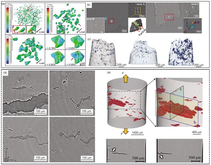
图5
同步辐射X射线原位成像技术在镁合金损伤断裂过程中的典型应用[67,70,71]
Fig.5
Typical applications of synchrotron radiation X-ray in situ imaging technology in the damage and fracture process of magnesium alloys
(a) von Mises stress (S) distributions on the particles in composites with various particle sizes at 0.8% strain and the fracture process of a representative particle[67] (ε—true strain. The stress concentration was indicated by red arrows)
(b, c) 2D slices (b) and 3D (c) fracture morphologies of composites[67] (White arrows in Fig.5b indicate microcracks. The blue regions in Fig.5c represent the microcracks)
(d) void/microcrack link-up via micro-scale localization in the surface-scans of the sample[70] (The three principal directions are labeled as longitudinal (L), which corresponds to the rolling direction, transverse (T) and short-transverse (S) through the thickness)
(e) reconstructed X-ray images showing cracks near the surface of the sample[71] (F—force. The arrow in the bottom left image of Fig.5e shows a shear-like linkage between cracks, while the arrow in bottom right image shows a deflected fatigue crack propagation path)
加载方式是影响镁合金损伤行为最重要的因素之一。Kondori等[70]利用同步辐射二维X射线原位成像技术揭示了退火态AZ31B镁合金的裂纹萌生与扩展模式(图5d[70])。结果表明,材料的损伤始于扁平孔洞,其钝化程度因空间位置、整体三轴度和局部应变水平而异。此外,实际使用的构件可能承受交变载荷,工程部件的疲劳寿命往往取决于材料在循环加载过程中的短裂纹状态[68]。Wang等[71]阐明了AZ31镁合金在单轴低周疲劳条件下主要变形过程与相关损伤机制之间的关系(图5e[71])。然而在实验环境方面,目前对材料变形行为的研究多在室温下进行,针对极端低温、高温、高速冲击等特殊工况的原位研究仍较为缺乏。在加载条件方面,为更贴合实际服役情况,尚需进一步研究交变载荷(剪切、扭转及复合载荷)下的损伤机制[72]。除环境因素外,镁合金内部的第二相、孪晶、晶粒尺寸等微观结构也对材料的损伤断裂行为具有重要影响。Azghandi等[73]通过实验室X射线原位成像技术,比较了晶粒尺寸分别约为3和60 μm的AZ31镁合金在不同应变水平下断裂孔洞的形成和演化,发现孔洞数量密度与应变近似呈线性关系。
4 镁合金的腐蚀机理与表面防护研究
腐蚀是镁合金的典型失效模式之一,对其结构完整性和功能稳定性构成显著威胁[76]。在生物医用场景中,镁合金因体内腐蚀速率过快致使植入体过早失效,难以满足长期支撑需求。其腐蚀过程伴随H2与OH-的释放,引发局部碱性环境。腐蚀产物的累积不仅阻碍内膜组织愈合与再生,还会诱发炎症反应。此外,腐蚀导致的力学性能骤降进一步制约了其临床转化[77]。在工程应用领域,镁合金因较低的电极电位而在潮湿或腐蚀性介质中易遭侵蚀,尤其在含Cl-环境下更为显著。例如,海洋工程及混凝土结构中的硫酸盐还原菌与Cl-反应会加速其腐蚀进程,从而限制了其大规模工程应用。深入探究镁合金的腐蚀行为可揭示其失效机制,为开发高效防护策略、延长镁合金服役寿命提供理论支撑。而对其腐蚀产物动态演化规律的研究,则有助于指导其在特定环境中的合理选材和应用。因此,系统解析镁合金的腐蚀机理和动力学过程,对优化材料性能、拓展生物医学应用、降低维护成本及推动工程实践具有重要意义[78]。
现阶段,镁合金腐蚀行为和机理的研究主要依托光学或电子显微技术和电化学手段对腐蚀形貌进行监测。然而,传统二维表面方法难以实现三维乃至四维视角下的腐蚀形态可视化。由于Mg易与第二相引发微电偶腐蚀并形成复杂的三维微观结构(如穿透基体及内部的深蚀坑),若无法实现腐蚀演化的三维动态观测,则难以建立微观组织、结构与腐蚀行为的定量关联。为此,科研人员进行了大量的先进成像实验,如运用X-CT技术评估Mg在NaCl及人体体液中的局部腐蚀速率[79],采用X-CT定量分析AZ91镁合金中第二相与腐蚀坑的空间对应关系[80]。其中,Tian等[81]通过原位X-CT系统研究了AlLi、Alₓ(Gd, Y, Mn)及镁锡(Mg2Sn)等第二相对Mg-Li合金腐蚀行为的影响,发现腐蚀坑由连通性良好的棒状深坑与枝晶状裂纹构成,且腐蚀前期受颗粒状AlLi相主导,后期则受块状Alₓ相和层状相调控(图6a[81])。然而,X-CT虽能提供静态三维结构信息,却受限于扫描速率与时间分辨率,难以捕捉快速腐蚀反应的动态过程,且侧重于已形成缺陷的分析而非演化机制研究。相较之下,同步辐射动态成像技术凭借其较高的时空分辨率优势,能够实时追踪镁合金在加工或服役环境中微观结构的动态演变(如晶界迁移及裂纹扩展等)。王同敏团队[82]采用同步辐射原位动态成像技术深入研究了Mg-Sn合金的腐蚀动力学过程,构建了腐蚀随时间演变的完整图谱。如图6b[82]所示,初始阶段于晶粒内部萌发腐蚀核,继而发展为坑蚀形态;随着腐蚀进程推进,蚀坑沿晶粒纵深扩展,并在晶界处受阻后转向横向生长,最终实现相邻晶粒间蚀坑的融合。此外,王同敏团队[83]还通过同步辐射四维成像技术原位观察LZ91镁锂合金在3.5%NaCl溶液中的浸泡行为,发现大量β-Li相发生剥离的现象,证实该相为优先腐蚀区域[83]。
图6

图6
同步辐射成像技术在镁合金腐蚀防护领域的应用[81,82,85,86]
Fig.6
Applications of synchrotron imaging technology in the corrosion protection of magnesium alloys
(a) 3D reconstructed images before and after 360 h immersion in 3.5%NaCl (mass fraction) solution for LAZWMVT alloy (Mg-Li-Al-Zn-Y-Mn-Gd-Sn alloy) under in situ corrosion observation, with secondary phase distributions indicated (Green represents Alₓ(Gd, Y, Mn) phase, red denotes Mg2Sn phase, light blue marks AlLi phase, and yellow highlights corrosion pits)[81]
(b) time-dependent corrosion morphologies of Mg-Sn alloy in NaCl solution monitored via in situ corrosion observation, and schematic diagram and physical device of in situ corrosion imaging experiment at SSRF[82]
(c) 3D reconstructions of the interface between MAO-SSC coating on LA81 Mg-Li alloy surface obtained through in situ corrosion observation[85] (MAO—micro-arc oxidation, SSC—solid-like slippery composite coating)
(d) 3D reconstructions of simulated ZX50 and MAO implant sites showing visible bubble structures (in light blue) via in situ corrosion observation[86]
针对镁合金腐蚀防护需求,开发高性能涂层体系具有重要战略意义。微弧氧化(MAO)作为合金的重要表面处理工艺,其固有的高孔隙率特征严重制约了长期防腐效能。传统显微分析方法受限于二维投影特性,难以实现对涂层三维微观结构的精确表征。Chen等[84]利用X-CT系统量化了阳极氧化涂层(PEO)中微缺陷的尺寸分布、体积占比及空间构型,发现该类涂层在腐蚀介质作用下,微缺陷会随浸泡时间延长而发生显著形变与扩展,最终形成贯通性缺陷通道[84]。王同敏团队[85]采用同步辐射三维成像技术,精细重构了MAO涂层的界面结合状态,清晰观察到涂层内部及表面广泛分布着随机取向的微米级孔洞与裂纹网络。此类结构通过机械嵌合作用有效增强了界面结合强度(图6c[85])。在生物医用材料研究领域,研究人员为评估骨组织与种植体的界面稳定性及生物相容性,将裸ZK50合金和MAO处理的ZX50镁合金植入大鼠股骨模型,并运用同步辐射X射线四维成像技术解析了腐蚀降解过程对骨整合及组织响应的影响机制。研究[86]表明,MAO涂层在植入早期可有效抑制界面处的降解速率,显著提升植入体初始稳定性。待骨愈合完成后,该涂层又能适时发生可控降解。这种时序调控特性有助于降低继发性术后损伤风险,展现出良好的临床转化潜力(图6d[86])。然而,因同步辐射X射线成像技术辐射剂量较高,不适用于长期活体动物实验;且无法区分生物组织与腐蚀产物的信号叠加,导致降解动力学参数测量误差较大。
5 总结和展望
在过去的数十年中,国内外研究人员基于同步辐射X射线进行了大量原位装置的建设,实现了凝固、变形及损伤断裂、腐蚀损伤的原位研究,为镁合金的微观结构演化提供了大量原位动态数据并进行了定量分析,解决了诸多镁合金制备过程和模拟服役环境下的基础科学问题。但客观而言,目前取得的这些科学成果只是众多基础科学问题中的一小部分,还有更多的关键科学问题有待进一步挖掘和解析。在未来,基于大科学装置开展镁合金的研究工作可在以下几方面给予重点关注。
(1) 为进一步优化研究策略,应注重同步辐射光源与实验室X射线装置的协同使用,发挥各自优势。例如,可先基于商业实验室X射线设备开展前期探索性实验,并用于原位装置的开发与验证,再利用同步辐射光源进行高精度动力学表征。对于需要长时间观察变化过程的实验(如长时生物植入实验过程、慢速腐蚀实验等),采用同步辐射或实验室光源的准原位实验则更为可行。
(2) 随着镁合金在极端高/低温、高压、电磁、腐蚀等多场耦合服役环境中的广泛应用,对其成分设计和制备工艺提出了更为精准的要求。因此,亟需推动面向复杂服役条件的样品制备与模拟环境装置的建设和发展。例如,对于镁合金氢气储能的应用需求,高压、高温环境下镁合金内部微结构转变的动力学机理仍有诸多科学问题需要探明,在同步辐射线站构建相关的高压、高温样品环境装置,利用同步辐射衍射等技术方法有望为研究微结构的转变过程提供新的视角。当前,针对镁合金在极端温度-复杂载荷(如拉伸、压缩、疲劳、扭转、剪切或其复合加载)耦合作用下的变形与断裂机理研究仍较为缺乏;在腐蚀介质与复杂受载协同环境中镁合金失效机制的原位评价也尚属空白;极端压力-温度耦合条件下的力学行为分析仍需深入。此外,模拟现代工业制造工艺(如电弧、激光选区增材制造)的原位实验装置,仍是未来重点发展的方向,以实现对真实工况的高保真复现与机理研究。
(3) 在镁合金研究中,以往的工作主要依赖同步辐射X射线照射样品,并采用单一探测器收集成像、衍射或能谱等某一类信号。为更全面揭示其微观机理,未来应大力推进多种X射线方法学的联用,例如,结合成像、衍射与微区光谱等技术,实现在单次实验中同步获取镁合金的微观形貌、物相分布、晶粒取向及成分信息。例如,可将白光Laue微衍射与微区荧光光谱联用,在开展微区衍射实验的同时,利用荧光探测器识别镁合金中非均匀分布的微量组分,从而实现对制备过程中成分与晶体组织变化的协同表征。同时,超快动力学实验(如每秒数十万帧的高速成像)在单次运行中即可产生海量数据,这对数据的实时传输、高效存储及自动化处理提出了极高要求。因此,亟需发展相应的快速传输与存储技术,并加强实验数据的自动提取、转换和智能分析算法的开发,推动高效、便捷的数据处理软件工具的研制。此外,将同步辐射与中子源等大型实验平台进行联动,也是在更大尺度范围内开展镁合金微区结构和内应力测量的重要发展方向。
参考文献
Towards development of a high-strength stainless Mg alloy with Al-assisted growth of passive film
[J].
Magnesium alloys with high strength and excellent corrosion resistance are always sought-after in light-weighting structural components for automotive and aerospace applications. However, for most magnesium alloys that have a high specific strength, they usually have an inferior corrosion resistance and vice versa. In this work, we successfully develop a Mg-11Y-1Al (wt. %) alloy through conventional casting, solution treatment followed by extrusion. The overall properties of this alloy feature with a corrosion rate lower than 0.2 mm y, high yield strength of 350 MPa and moderate tensile elongation of 8%, the combination of which shows competitive advantage over other comparative magnesium alloys in the literature. It is found that a thin and dense protective film of YO/Y(OH) can be fast developed with the aid of AlO/Al(OH) deposition to isolate this alloy from further attack of corrosion medium. Meanwhile, the refined grains, weak texture and activation of non-basal slip systems co-contribute to the high strength and good ductility. Our findings are expected to inspire the design of next-generation high performance magnesium alloys.© 2022. The Author(s).
Anomalous strain hardening via manipulating basal/pyramidal dislocation interactions in the Mg-Y-Ca alloy
[J].
Machine learning pipeline for structure-property modeling in Mg-alloys using microstructure and texture descriptors
[J].
The effect of slow shot speed and casting pressure on the 3D microstructure of high pressure die casting AE44 magnesium alloy
[J].
The evolution of the growth morphology in Mg-Al alloys depending on the cooling rate during solidification
[J].
Temperature-dependent interplay of intra- and inter-granular deformation mechanisms in Mg-10Y: Statistical analysis from an HRDIC perspective
[J].
In-situ TEM observation of
Magnetic modulation of keyhole instability during laser welding and additive manufacturing
[J].Keyhole instability during laser welding and laser powder bed fusion (LPBF) can cause keyhole collapse and pore formation. Using high-speed x-ray imaging, we demonstrate that the flow vortex-induced protrusion on the rear keyhole wall is crucial in initiating keyhole instability. Applying a transverse magnetic field suppresses the keyhole instability by driving a secondary thermoelectric magnetohydrodynamics (TEMHD) flow that alters the net flow vortex. This minimizes protrusions and large-amplitude keyhole oscillations. The suppression effectiveness depends on the laser scanning direction relative to the magnetic field orientation because this controls the Seebeck effect-induced Lorentz force's direction. We show that at LPBF length scales, electromagnetic damping is weak, and for alloys with a large Seebeck coefficient, TEMHD becomes the dominant mechanism controlling flow behind the keyhole.
Characterization of just one atom using synchrotron X-rays
[J].
Development of magnesium alloys: Advanced characterization using synchrotron radiation techniques
[J].Magnesium alloys are the lightest metal structural materials owing to their excellent physical and chemical properties. Microstructural evolution in magnesium alloys under the conditions of casting, thermal-mechanical processing, and in-service environment, play an important role in governing their mechanical properties and reliability/sustainability. A synchrotron light source produces high flux, tunable X-ray energy, high resolution, and high coherence X-ray beams, which can realize <em>in-situ</em> dynamic observation of microstructural evolution in a wide range of alloys during the entire processing chain and in simulated service environments. This article reviews the fundamentals of synchrotron radiation characterization techniques (imaging, diffraction, scattering, and fluorescence holography) and state-of-the-art advanced synchrotron characterization techniques on the microstructure evolution mechanism of magnesium alloys. Case studies span a broad range of solidification, deformation, precipitation, fracture and damage, corrosion, and energy storage. Research opportunities and challenges of physical metallurgy studies of magnesium alloys are highlighted for future studies.
Time resolved X-ray imaging of dendritic growth in binary alloys
[J].
Influence of growth velocity on fragmentation during directional solidification of Al-14 wt.% Sn alloy studied by in-situ synchrotron X-radiography
[J].
Revealing dendritic pattern formation in Ni, Fe and Co alloys using synchrotron tomography
[J].
In situ experiments with X ray tomography: An attractive tool for experimental mechanics
[J].
Real-time quantitative imaging of failure events in materials under load at temperatures above 1, 600 oC
[J].
Unravelling dynamic recrystallisation in a microalloyed steel during rapid high temperature deformation using synchrotron X-rays
[J].
Study on diffusion behavior and microstructural evolution of Al/Cu bimetal interface by synchrotron X-ray radiography
[J].
Evolution of dendrite morphology of a binary alloy under an applied electric current: An in situ observation
[J].
The effect of anisotropic microstructure on the crack growth and fatigue overload behaviour of ultrafine-grained nickel
[J].Changes in crack growth rate associated with overload events during fatigue are poorly understood, especially for materials with anisotropic microstructures. Here overload fatigue tests are reported for compact tension samples cut in two different orientations from high pressure torsion disc samples. During growth the crack planes reoriented either slightly, or significantly, to align with the elongated grain structure leading to low, and high, levels of mixed mode fatigue loading respectively. In both cases the ultrafine grained microstructure led to macroscopically flat crack faces. The fatigue crack growth rate was around 2.4 times slower for the case with the high mode II component than for the low. A 100% overload was then introduced and synchrotron X-ray diffraction and digital image correlation (DIC) were applied in-itu to map the bulk crack-tip elastic strain field (plane strain) and surface displacement field (plane stress) respectively prior to, during and after overload. The high mode II case displayed a larger degree of retardation after overload. Residual stress and plasticity-induced crack closure were found to be the primary causes for the retardation as the crack grows into the overload plastic zone. Significant crack face contact was observed for the high mode II case along with significant levels of compressive stress transferred across the crack faces at minimum load. Compared with conventional (coarse) grain Ni, the ultrafine grained Ni is less retarded by overload, because of its relatively flatter crack path and higher yield stress and thus less plasticity and residual stress induced closure. (C) 2019 Acta Materialia Inc. Published by Elsevier Ltd.
Critical damage events of 3D printed AlSi10Mg alloy via in situ synchrotron X-ray tomography
[J].
Structural integrity issues of composite materials and structures in future transportation equipment
[J].
α-Mg primary phase formation and dendritic morphology transition in solidification of a Mg-Nd-Gd-Zn-Zr casting alloy
[J].
Effect of additional solute elements (X = Al, Ca, Y, Ba, Sn, Gd and Zn) oncrystallographic anisotropy during the dendritic growth of magnesium alloys
[J].
Intermittent nucleation and periodic growth of grains under thermo-solutal convection during directional solidification of Al-Cu alloy
[J].
Phase-field simulations of dendrite morphologies and selected evolution of primary α-Mg phases during the solidification of Mg-rich Mg-Al-based alloys
[J].
Characterisation of the 3-D dendrite morphology of magnesium alloys using synchrotron X-ray tomography and 3-D phase-field modelling
[J].
Effect of different solute additions on dendrite morphology and orientation selection in cast binary magnesium alloys
[J].
Synchrotron tomographic quantification of the influence of Zn concentration on dendritic growth in Mg-Zn alloys
[J].
Fast synchrotron X-ray tomographic quantification of dendrite evolution during the solidification of Mg Sn alloys
[J].
Dendritic evolution during coarsening of Mg-Zn alloys via 4D synchrotron tomography
[J].
Universality and self-similarity in pinch-off of rods by bulk diffusion
[J].
The influence of SiC-Al2O3 reinforcements on the deformation and fracture mechanism of thixomolded Mg-based composite
[J].
The effect of micro-SiCp content on the tensile and fatigue behavior of AZ61 magnesium alloy matrix composites
[J].
Simultaneously improving the strength and ductility of AZ91/GNPs composites through decorating graphene nanoplatelets with MgO
[J].Magnesium matrix composites (MgMCs) have always suffered low strengthening efficiency and poor ductility due to the difficulties in pursuing the well-bonded interface. Herein, graphene nanoplatelets (GNPs) were decorated with magnesium oxide nanoparticles (MgO NPs) through chemical co-precipitation and then incorporated into AZ91 alloy to fabricate MgMCs via powder thixoforging. The effect of MgO on the interface of the Mg/graphene system was investigated based on the first-principles calculations, and the result indicated that modifying GNPs with MgO NPs was helpful in improving the Mg-GNP interface bonding. The interface structural analysis revealed that the MgO NPs were firmly bonded with both GNPs and α-Mg through the distortion area bonding and semi-coherent interfacial bonding, severing as a bridge to fasten the interface bonding of composites. In addition, the MgO NPs on GNPs acted as a barrier to prevent GNPs from seriously reacting with the AZ91 alloy. As a result, the AZ91/MgO@GNPs composite was endowed with enhancements of 31% and 10% in the yield strength, and increments of 71% and 61% in elongation compared with the AZ91 alloy and AZ91/GNPs composite, respectively, exhibiting a more significant potential in optimizing the strength-toughness tradeoff compared with the AZ91/GNPs. Moreover, the possible strengthening and toughening mechanisms were also discussed in detail. This work offers a relatively novel surface modification strategy to modulate the Mg-GNP interface for a simultaneous improvement of strength and ductility.
Grain refinement mechanism of SiC nanoparticles/Mg-9 wt.% Al composite investigated by sharp interface model at microscale and nanoscale
[J].
The influence of nanoparticles on dendritic grain growth in Mg alloys
[J].
Unraveling the individual and synergistic effects of copper and gadolinium on the microstructure and mechanical properties of Mg-6Zn-0.5Ca alloy
[J].
A synergetic approach to enhancing mechanical properties of trace TiB2 particles reinforced AZ91D composites through α-Mg grain refinement and β-Mg17Al12 manipulation
[J].
Achieving simultaneous refinement of α-Mg grains and β-Mg17Al12 phases via trace multi-scale TiB2 for strength-ductility synergy in AZ91D composites
[J].
Grain refinement and mechanical property enhancement of AM60B magnesium alloy via TiB2 particle regulation
[J].
The effect of slow shot speed and casting pressure on the 3D microstructure of high pressure die casting AE44 magnesium alloy
[J].
Quantum optics: Science and technology in a new light
[J].Light facilitates exploration of quantum phenomena that illuminate the basic properties of nature and also enables radical new technologies based on these phenomena. The critical features of quantum light that underpin the opportunities for discovery and application are exceptionally low noise and strong correlations. Rapid progress in both science and technology has been stimulated by adopting components developed for optical telecommunications and networking, such as highly efficient detectors, integrated photonic circuits, and waveguide- or nanostructure-based nonlinear optical devices. These provide the means to generate new quantum states of light and matter of unprecedented scale, containing many photons with quantum correlations across space and time. Notably, networks with only several tens of photons are already beyond what can be efficiently analyzed by current computers. Copyright © 2015, American Association for the Advancement of Science.
Designing a magnesium alloy with high strength and high formability
[J].Although magnesium alloys, as the lightest structural alloys, offer significant potential for automotive applications, their applications remain limited due to their poor formability at room temperature. Since the strategies used for improving formability usually result in a degradation of strength, there are no high strength magnesium alloys showing good formability. Here we report an alloy design concept that can simultaneously provide high strength and good formability. Such designed alloy when subjected to an appropriate processing technique shows a combination of strength and formability that surpasses those of the existing magnesium alloys reported so far. The alloy design concept used in the present study is based on the utilization of alloying elements that can induce precipitation, as well as maximize the segregation of other texture-controlling alloying elements. Such developed alloy is expected to broaden the application of Mg alloy sheets, which are now starting to gain acceptance by automotive industries.
Grain nucleation and growth during phase transformations
[J].The mechanical properties of polycrystalline materials are largely determined by the kinetics of the phase transformations during the production process. Progress in x-ray diffraction instrumentation at synchrotron sources has created an opportunity to study the transformation kinetics at the level of individual grains. Our measurements show that the activation energy for grain nucleation is at least two orders of magnitude smaller than that predicted by thermodynamic models. The observed growth curves of the newly formed grains confirm the parabolic growth model but also show three fundamentally different types of growth. Insight into the grain nucleation and growth mechanisms during phase transformations contributes to the development of materials with optimal mechanical properties.
High-pressure strengthening in ultrafine-grained metals
[J].
In situ observation of crystal rotation in Ni-based superalloy during additive manufacturing process
[J].Understanding the dynamic process of epitaxial microstructure forming in laser additive manufacturing is very important for achieving products with a single crystalline texture. Here, we perform in situ, real-time synchrotron Laue diffraction experiments to capture the microstructural evolution of nickel-based single-crystal superalloys during the rapid laser remelting process. In situ synchrotron radiation Laue diffraction characterises the crystal rotation behaviour and stray grain formation process. With a complementary thermomechanical coupled finite element simulation and molecular dynamics simulation, we identify that the crystal rotation is governed by the localised heating/cooling heterogeneity-induced deformation gradient and recognise that the sub-grain rotation caused by rapid dislocation movement could be the origin of granular stray grains at the bottom of the melt pool.© 2023. The Author(s).
Watching the growth of bulk grains during recrystallization of deformed metals
[J].We observed the in situ growth of a grain during recrystallization in the bulk of a deformed sample. We used the three-dimensional x-ray diffraction microscope located at the European Synchrotron Radiation Facility in Grenoble, France. The results showed a very heterogeneous growth pattern, contradicting the classical assumption of smooth and spherical growth of new grains during recrystallization. This type of in situ bulk measurement opens up the possibility of obtaining experimental data on scientific topics that before could only be analyzed theoretically on the basis of the statistical characterization of microstructures. For recrystallization, the in situ method includes direct measurements of nucleation and boundary migration through a deformed matrix.
Measuring the stress field around an evolving crack in tensile deformed Mg AZ31 using three-dimensional X-ray diffraction
[J].
Observation of non-basal slip in Mg-Y by in situ three-dimensional X-ray diffraction
[J].
Onset of detwinning in Mg-3Al-1Zn alloy: A synchrotron-based X-ray diffraction study
[J].
Deformation and texture evolution in AZ31 magnesium alloy during uniaxial loading
[J].
Local lattice distortion mediated formation of stacking faults in Mg alloys
[J].
Plasticity analysis by synchrotron radiation in a Mg97Y2Zn1 alloy with bimodal grain structure and containing LPSO phase
[J].
The effect of temperature on load partitioning evolution in magnesium metal matrix composite reinforced with Ti particles using in-situ synchrotron radiation diffraction experiments
[J].
Deformation twinning and grain partitioning in a hexagonal close-packed magnesium alloy
[J].Pervasive deformation twinning in magnesium greatly affects its strength and formability. The local stress fields associated with twinning play a key role on deformation behavior and fracture but are extremely difficult to characterize experimentally. In this study, we perform synchrotron experiments with differential-aperture X-ray microscopy to measure the 3D stress fields in the vicinity of a twin with a spatial resolution of 0.5 micrometer. The measured local stress field aids to identify the sequence of events involved with twinning. We find that the selected grain deforms elastically before twinning, and the twin formation splits the grain into two non-interacting domains. Under further straining one domain of the grain continued to deform elastically, whereas the other domain deforms plastically by prismatic slip. This heterogeneous deformation behavior may be mediated by the surrounding medium and it is likely to lead to asymmetric twin growth.
Mechanism-based constitutive modeling of ZEK100 magnesium alloy with crystal plasticity and in-situ HEXRD experiment
[J].
Multi-scale mechanisms of twinning-detwinning in magnesium alloy AZ31B simulated by crystal plasticity modeling and validated via in situ synchrotron XRD and in situ SEM-EBSD
[J].
Studying the effect of stress relaxation and creep on lattice strain evolution of stainless steel under tension
[J].
Individual phase constitutive properties of a TRIP-assisted QP980 steel from a combined synchrotron X-ray diffraction and crystal plasticity approach
[J].
An in situ high-energy X-ray diffraction study of micromechanical behavior of multiple phases in advanced high-strength steels
[J].
Incorporation of twinning into a crystal plasticity finite element model: Evolution of lattice strains and texture in Zircaloy-2
[J].
In-situ neutron diffraction of a quasicrystal-containing Mg alloy interpreted using a new polycrystal plasticity model of hardening due to {10.2} tensile twinning
[J].
In situ neutron diffraction and polycrystal plasticity modeling of a Mg-Y-Nd-Zr alloy: Effects of precipitation on individual deformation mechanisms
[J].
Modeling lattice strain evolution at finite strains and experimental verification for copper and stainless steel using in situ neutron diffraction
[J].
Strain rate sensitivities of deformation mechanisms in magnesium alloys
[J].
Three-dimensional nucleation and growth of deformation twins in magnesium
[J].At two-thirds the weight of aluminum, magnesium alloys have the potential to reduce the fuel consumption of transportation vehicles. These advancements depend on our ability to optimize the desirable versus undesirable effects of deformation twins, which are three-dimensional (3D) microstructural domains that form under mechanical stresses. Previously only characterized through surface or thin-film measurements, we present 3D in situ characterization of deformation twinning inside an embedded grain over mesoscopic fields of view using dark-field x-ray microscopy supported by crystal plasticity finite element analysis. The results revealed the role of triple junctions on twin nucleation and the sequence and irregularity of twin growth and showed that twin-grain junctions, twin-twin junctions, and twin boundaries were the sites of localized dislocation accumulation.
Mechanical behavior of SiC reinforced ZA63 Mg matrix composites: Experiments and 3D finite element modelling
[J].
Inhibiting weld cracking in high-strength aluminium alloys
[J].Cracking from a fine equiaxed zone (FQZ), often just tens of microns across, plagues the welding of 7000 series aluminum alloys. Using a multiscale correlative methodology, from the millimeter scale to the nanoscale, we shed light on the strengthening mechanisms and the resulting intergranular failure at the FQZ. We show that intergranular AlCuMg phases give rise to cracking by micro-void nucleation and subsequent link-up due to the plastic incompatibility between the hard phases and soft (low precipitate density) grain interiors in the FQZ. To mitigate this, we propose a hybrid welding strategy exploiting laser beam oscillation and a pulsed magnetic field. This achieves a wavy and interrupted FQZ along with a higher precipitate density, thereby considerably increasing tensile strength over conventionally hybrid welded butt joints, and even friction stir welds.© 2022. The Author(s).
A new strategy for fabricating Mg-Al alloys with excellent strength-ductility synergy via pulse-coupled wire-arc directed energy deposition
[J].
Void growth and coalescence in a magnesium alloy studied by synchrotron radiation laminography
[J].
In-situ synchrotron X-ray tomography investigation of damage mechanism of an extruded magnesium alloy in uniaxial low-cycle fatigue with ratchetting
[J].
Transgranular liquation cracking of grains in the semi-solid state
[J].Karagadde, S.; Lee, P. D.; Cai, B.; Azeem, M. A.; Tsivoulas, D. Univ Manchester, Sch Mat, Manchester Xray Imaging Facil, Manchester M13 9PL, Lancs, England. Karagadde, S.; Lee, P. D.; Cai, B.; Azeem, M. A. Res Complex Harwell, Didcot OX11 0FA, Oxon, England. Fife, J. L. Paul Scherrer Inst, Swiss Light Source, CH-5232 Villigen, Switzerland. Kareh, K. M. Univ London Imperial Coll Sci Technol & Med, Dept Mat, London SW7 2AZ, England. Puncreobutr, C. Chulalongkorn Univ, Dept Met Engn, Bangkok 10330, Thailand. Connolley, T.; Atwood, R. C. Diamond Light Source Ltd, Didcot OX11 0DE, Oxon, England.
A rationale for the influence of grain size on failure of magnesium alloy AZ31: An in situ X-ray microtomography study
[J].
Elementary growth mechanisms of creep cavities in AZ31 alloy revealed by in situ X-ray nano-tomography
[J].
Nanotomographic evaluation of precipitate structure evolution in a Mg-Zn-Zr alloy during plastic deformation
[J].Magnesium and its alloys attract increasingly wide attention in various fields, ranging from transport to medical solutions, due to their outstanding structural and degradation properties. These properties can be tailored through alloying and thermo-mechanical processing, which is often complex and multi-step, thus requiring in-depth analysis. In this work, we demonstrate the capability of synchrotron-based nanotomographic X-ray imaging methods, namely holotomography and transmission X-ray microscopy, for the quantitative 3D analysis of the evolution of intermetallic precipitate (particle) morphology and distribution in magnesium alloy Mg-5.78Zn-0.44Zr subjected to a complex multi-step processing. A rich history of variation of the intermetallic particle structure in the processed alloy provided a testbed for challenging the analytical capabilities of the imaging modalities studied. The main features of the evolving precipitate structure revealed earlier by traditional light and electron microscopy methods were confirmed by the 3D techniques of synchrotron-based X-ray imaging. We further demonstrated that synchrotron-based X-ray imaging enabled uncovering finer details of the variation of particle morphology and number density at various stages of processing-above and beyond the information provided by visible light and electron microscopy.
In vitro dynamic degradation behavior of new magnesium alloy for orthopedic applications
[J].
Fundamentals and advances in magnesium alloy corrosion
[J].
Assessment of localized corrosion under simulated physiological conditions of magnesium samples with heterogeneous microstructure: Value of X-ray computed micro-tomography platform
[J].
Quantifying the influence of secondary phases on corrosion in multicomponent Mg alloys using X-ray computed microtomography
[J].
Quantifying the influence of microstructure on the corrosion of Mg-Li alloys by using X-ray CT
[J].
Corrosion process of Mg-Sn alloys revealed via in situ synchrotron X-ray radiography
[J].
Influence of deformation on the corrosion behavior of LZ91 Mg-Li alloy
[J].
3-D distribution characteristics of the micro-defects in the PEO coating on ZM6 Mg-alloy during corrosion
[J].
Mao-based solid-like slippery composite coating with superior corrosion resistance and robust machinery performance upon magnesium-lithium alloy LA81
[J].
In vivo degradation performance of micro-arc-oxidized magnesium implants: A micro-CT study in rats
[J].Biodegradable Mg alloys are of great interest for osteosynthetic applications because they do not require surgical removal after they have served their purpose. In this study, fast-degrading ZX50 Mg-based implants were surface-treated by micro-arc oxidation (MAO), to alter the initial degradation, and implanted along with untreated ZX50 controls in the femoral legs of 20 male Sprague-Dawley rats. Their degradation was monitored by microfocus computed tomography (μCT) over a total observation period of 24weeks, and histological analysis was performed after 4, 12 and 24weeks. While the MAO-treated samples showed almost no corrosion in the first week, they revealed an accelerated degradation rate after the third week, even faster than that of the untreated ZX50 implants. This increase in degradation rate can be explained by an increase in the surface-area-to-volume ratio of MAO-treated implants, which degrade inhomogeneously via localized corrosion attacks. The histological analyses show that the initially improved corrosion resistance of the MAO implants has a positive effect on bone and tissue response: The reduced hydrogen evolution (due to reduced corrosion) makes possible increased osteoblast apposition from the very beginning, thus generating a stable bone-implant interface. As such, MAO treatment appears to be very interesting for osteosynthetic implant applications, as it delays implant degradation immediately after implantation, enhances fracture stabilization, minimizes the burden on the postoperatively irritated surrounding tissue and generates good bone-implant connections, followed by accelerated degradation in the later stage of bone healing.Copyright © 2012 Acta Materialia Inc. Published by Elsevier Ltd. All rights reserved.
Superhydrophobic and corrosion-resistant coating on magnesium-lithium alloys
[J].
Boosting corrosion resistance of Mg-Li alloy: Implanting bioinspired superhydrophobic surfaces into MAO matrix for enhanced protection
[J].
MAO-based self-healing phase-change solid-like slippery composite coating with superior corrosion resistance and robust machinery performance
[J].Super-slippery surfaces have exhibited significant promise for corrosion protection of metals. However, challenges remain in the way of broad engineering applications, including limited mechanical durability, conflict between mechanical performance and self-healing ability, and volatilization and leakage of lubricant caused by long-term usage. This study aims to improve mechanical strength and minimize liquid loss of super-slippery surfaces by using a silane coupling agent (KH550) as bridging agent to bind phase-change paraffin wax, hydrophilic nano-silica, and low-surface-energy polydimethylsiloxane (PDMS) onto epoxy-functionalized surfaces via a series of dehydration, condensation, and cross-linking reactions. This method yields a super-slippery, corrosion-resistant composite coating with phase-change self-healing properties on magnesium-lithium alloy LA81. It demonstrates a high water-contact angle (> 110°) and a low sliding angle (< 8°), indicative of favorable hydrophobic and slippery characteristics. Electrochemical tests reveal a profound increment (seven orders of magnitude) in impedance modulus, substantiating its enhanced anti-corrosive performance. In addition, coating integrity was maintained after 168 h of salt spray exposure. Adhesion tests and 3D synchrotron X-ray imaging confirm a strong bond between the coating and substrate. Due to paraffin’s phase-change properties, the composite coating exhibits rapid self-healing when thermally stimulated. Such a self-cleaning, corrosion-resistant, and self-healing composite coating, with exceptional mechanical properties, offers an alternative solution to extending material lifespans in engineering applications.


