


纳米多孔金属是一种具有双连续结构的材料,因其较高的孔隙率和比表面积,在催化[1,2]、驱动器[3,4]、传感器[5,6]及电池[7]领域具有广泛的应用。通常采用脱合金法制备纳米多孔金属,将合金中活性较高的成分溶解分离出去,留下相对惰性的合金元素,最终形成一种海绵状的纳米多孔材料[8,9]。例如,在Au-Ag合金中有选择性地将Ag溶解后,会形成由许多孔棱和孔洞组成的纳米多孔Au[10]。通过控制脱合金过程中的参数可以调节孔棱及孔洞的特征尺寸,使其在几纳米到几百纳米的范围内变化。由于尺寸效应等作用,往往通过降低孔棱的尺寸可以获取优异的性能。然而随着孔棱尺寸的降低,比表面积进一步升高,纳米多孔金属的孔棱在表面能的驱动下会发生粗化现象,影响材料的催化及力学性能[11]。
纳米多孔金属的表面结构和成分可以显著地影响其稳定性。纳米多孔Au的粗化可以理解为一种曲率驱动的生长,孔棱表面的Au原子从正曲率的凸面(通常为孔棱中心区域)向着负曲率的凹面扩散[12]。粗化的最终结果是孔棱在颈部断裂,其过程类似于纳米线的Rayleigh失稳[13]。为控制孔棱尺寸,防止其粗化,可以降低脱合金温度[14]或者在孔棱表面覆盖一层氧化物[15]。有研究[16]表明,在α黄铜中添加少量的As 可以有效抑制Cu的表面扩散,进而降低Zn的脱落,受此启发,Snyder等[17]在Au-Ag合金中添加了约6% (原子分数,下同)的Pt,发现孔棱粗化程度显著降低。基于Erlebacher[18]的孔隙演化模型,Snyder等[17]认为由于Pt的表面扩散速率比Au慢很多,Pt分离出合金后会富集在表面特别是台阶边缘位置,可以有效控制Au富集的小岛长大,从而抑制粗化。虽然这个假设有一定的合理性,但是尚未有直观的证据支持这一观点。Vega和Newman[19]通过在Au-Ag合金中添加不同比例的Pt发现,随着Pt的比例由1%增加到3%,孔棱的平均尺寸由6.8 nm降到了4.3 nm。此外他们[20]还研究了有氧和无氧环境下合金表面Pt的覆盖率,发现由于O和Pt之间强烈的相互作用,O有助于Pt钉扎在孔棱表面,O和Pt的共偏析会阻碍纳米多孔Au的粗化。不仅如此,他们[21]还发现Pt与Au协同作用可以提高纳米多孔金属对CH3OH氧化反应的催化性能。El-Zoka等[22]利用原子探针层析(APT)技术研究了Pt防止孔棱粗化的机理,通过半定量分析发现孔棱细化不仅与Pt对表面扩散的抑制有关,还取决于Ag在孔棱中的溶解程度。遗憾的是,虽然他们观察到了Pt在孔棱表面的偏聚,但是未能观察到Pt具体偏聚的位置。Cai等[23]认为孔棱表面低配位位点的原子更易溶解,而Pt会偏聚在这些位置阻碍孔棱粗化。虽然关于Pt防止孔棱粗化的观点很多,但迄今为止都未有直观的证据能够证明Pt的作用。为解决此问题,需要在原子尺度探究纳米多孔金属的表面结构及元素分布。
扫描电子显微镜(SEM)常用于表征材料的表面特征[24],但是其分辨率远远未到原子尺度。与之相比,扫描隧道显微镜(STM)的分辨率能够达到原子尺度[25,26],然而由于其仅探测表面1~2层原子且对样品平整度具有较高要求,难以对孔隙较多、表面起伏很大、呈海绵状的纳米多孔金属进行表征。APT技术能在纳米尺度获得样品的三维(3D)成分信息[27,28],但其未能将成分与原子位置真正对应起来,即难以精确定位元素偏聚的具体位置。高分辨透射电子显微镜(TEM)和高分辨扫描透射电子显微镜(STEM)是目前研究晶体原子结构的常用手段[29~34]。然而,由于这些技术只是获得二维(2D)投影图像,通常不能直接定量获取纳米晶体的3D结构,特别是它们的表面晶体学和配位信息[35],甚至有时候会产生误导。与传统的TEM和STEM成像方法相比,原子分辨率的电子层析(AET)技术[35~38]提供了一种解析材料的3D原子级信息的有力手段。本工作结合STEM和AET技术对纳米多孔Au的微观结构进行了表征,在原子尺度绘制了孔棱表面的配位环境。通过将AET技术与能量色散谱(EDS)成分分析结合,探究了Pt在纳米多孔Au-Pt表面的偏聚,以期为研究纳米多孔金属材料的结构和成分信息提供有力的技术方法支撑。
1 实验方法
制备纳米多孔金属的母合金分别为Au25Ag75及(Au0.95Pt0.05)25Ag75 (原子分数,%)。分别将纯度大于99.99% (质量分数)的Au、Ag和Pt金属丝依据各自配比计算出所需的质量。配比好对应母合金成分后,将其放入真空电弧炉中进行电弧熔炼。在合金熔炼前利用分子泵将真空抽至5 × 10⁻³ Pa,之后充入高纯的Ar气进行保护。每次熔炼结束将合金锭翻转后再次熔炼,重复7~8次以确保合金成分均匀。对于含Pt的合金样品,在多次熔炼的基础上逐步稀释熔炼。将熔炼后的合金放入真空石英管中在900℃均匀化退火100 h。均匀化处理后的合金薄片先进行线切割,然后减薄成直径3 mm、厚30 μm的金属薄片。当金属薄片出现凹坑后,在Gatan Model 691精密离子刻蚀系统上对其离子研磨直至薄片中间穿孔,制备出TEM样品。利用固相脱合金法[38]对样品进行脱合金,得到的样品后续用于TEM表征。
TEM及电子层析实验在像差校正Titan Cubed Themis G2 60-300型TEM上进行,加速电压为300 kV。电子层析实验采集高角环形暗场(HAADF)像时电子束的会聚半角为0.0249 rad,相机常数为90 mm,样品杆为Fischione 2020重构杆。电子层析实验的HAADF像大小为53.45 nm × 53.45 nm。像差校正电镜配备了Super X EDS系统用于获取EDS。在±75°的角度范围内间隔2°倾转样品,获取每一角度的HAADF像和EDS。对所有HAADF像及EDS进行降噪等图像处理后,利用同步迭代重构技术(simultaneous iterative reconstruction technique)对其重构获得物体的3D结果,原子识别及3D定量分析通过编写代码实现。
2 实验结果
2.1 纳米多孔Au的微观结构
图1
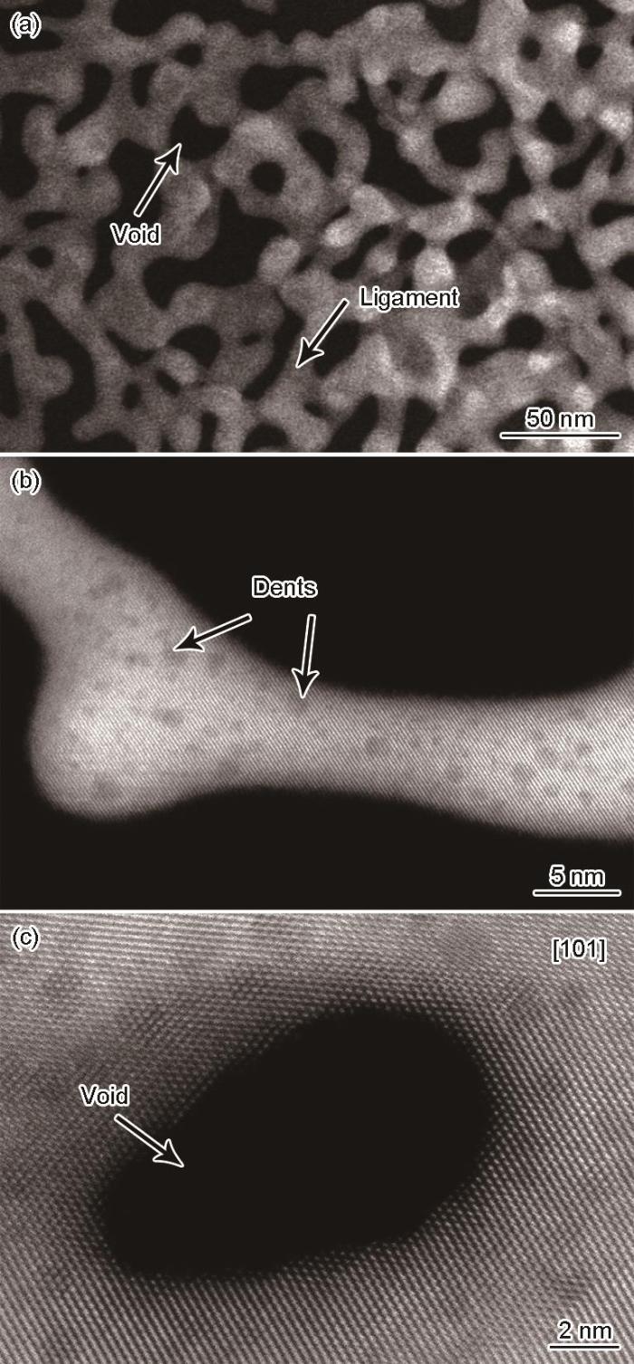
图1
纳米多孔Au的孔棱和孔隙结构高角环形暗场(HAADF)像
Fig.1
Low (a) and high (b, c) magnified high-angle annular dark field (HAADF) images of ligaments and voids in nanoporous gold
2.2 纳米多孔Au的三维结构与成分分布
图2

图2
低倍下纳米多孔Au结构和成分的三维(3D)重构结果
Fig.2
3D reconstructions of nanoporous gold structure (a) and chemical composition (b)
2.3 纳米多孔Au表面的缺陷结构
基于像差校正HAADF像,利用AET技术重构了直径约为10 nm孔棱的3D结构,如图3所示。图3a~c分别为该孔棱重构结果在[110]、[101]和[011] 3个晶带轴方向的投影。从不同方向的投影中都可以看到直径约2 nm的孔洞或凹坑。在每一个方向上的投影图中,原子都清晰可见。为判定重构结果的分辨率,对其进行了Fourier变换,从其功率谱(图3a中的插图)中测量出分辨率为0.12 nm。利用原子分辨率的3D重构结果对孔棱3D原子结构进行分析,通过寻峰获取每个原子坐标,计算出每个原子的配位数,由此绘制出孔棱表面原子的配位环境,如图3d所示。图3d中每个原子按照其配位数进行着色,例如配位数为9的(111)平台上的原子是紫红色,配位数为6的扭折和配位数为7的台阶分别为绿松石色和绿色。可以看到孔棱表面的缺陷可以大致分为2类,一类是(111)平台上的扭折和台阶,一类是表面凹坑。相比于扭折和台阶,表面凹坑会引入更多的低配位位点。
图3

图3
纳米多孔Au孔棱的原子分辨率电子层析3D重构及表面配位分析
Fig.3
Images of the reconstructed ligament with atomic resolution viewed along [110] (a), [101] (b), and [011] (c) zone axes, and the coordination map (d) (Inset in Fig.3a is the 3D power spectrum of the reconstruction showing a spatial resolution of 0.12 nm. The color bar on the right side of Fig.3d shows the corresponding rendered colors for atoms with different coordination numbers)
2.4 Pt元素的偏聚
为了探究Pt元素偏聚的具体位置,基于像差校正HAADF像,对纳米多孔Au-Pt的孔棱进行了电子层析3D重构,结果如图4所示。可见,该孔棱表面有许多大小不同的凹坑或孔洞。选取中间一个尺寸约为5 nm的孔洞,将原子分辨率电子层析与EDS的3D重构相结合来表征Pt元素的偏聚,结果如图5所示。图5a中孔洞的3D结果对应于图4的中间区域,孔洞边缘有许多低配位位点。图5b显示了Pt元素在孔棱上的3D分布,依据Pt元素EDS信号的强度对3D结果进行渲染(由蓝色到绿色逐步变强),将EDS渲染结果与3D原子结构叠加,可以看出Pt在孔棱表面的分布并不均匀。从3D结果的切片分析(图5c~f)来看,Pt集中分布在孔洞边缘低配位位点上。与此同时,观察到Pt既可以在{111}面这样的低指数晶面偏聚,也可以在{332}面这样的高指数晶面上偏聚。Au的{111}面表面能最低,{332}和{322}面次之,而{113}、{100}和{110}面表面能最高[39]。从图5c~f可以看出,Pt主要偏聚在{111}和{332}这样具有较低表面能的表面上,而在{113}这样表面能较高的面上则很少偏聚。
图4

图4
纳米多孔Au-Pt表面凹坑的原子级3D重构结果
Fig.4
3D atomic structure of surface dents in nanoporous Au-Pt
图5

图5
纳米多孔Au-Pt表面偏聚的3D分析
Fig.5
3D analyses of surface segregation in nanoporrous Au-Pt (The thickness of the slices is 1 nm)
(a) 3D reconstruction of the void in the surface
(b) 3D reconstruction of Pt distribution around the void in Fig.5a (The color represents the intensity of the EDS signal of the Pt element, which gradually becomes more intense from blue to green)
(c-f) multiple slices of 3D reconstructed chemical composition distribution on the surface of the void in Fig.5a (Crystallog-raphic planes with different indexes are determined and indicated)
3 分析讨论
3.1 孔棱表面的低配位位点与Pt的偏聚
脱合金法制备纳米多孔金属主要涉及2个步骤:一是Ag从母合金中溶解分离出去,形成初始的纳米多孔结构;二是随着孔棱的生长和粗化,残余的Ag进一步从纳米多孔结构内析出[40,41]。在固相脱合金过程中,首先利用Ar和O2的等离子体将Ag氧化使得孔棱表面覆盖AgO x (x在0.5~1.5之间),在等离子体的溅射作用下AgO x 层剥落,Ag进一步由内部扩散出去,形成初始的纳米多孔结构;然后在氩离子的刻蚀作用下,破坏氧化层,促进内部残余Ag的析出[38]。结合理论模型[8]和孔棱表面的3D原子结构可以推出,当Ag原子从母合金内分离出去后,会在原来的位置留下空位,附近的Ag原子因配位数降低也会进一步溶解,最终结果是空位沿着{111}面或{020}面扩散。随着Ag原子的减少,这些空位在扩散的同时还会形成空位簇(凹坑),剩余的Au原子也会沿着表面朝着{111}面或{020}面的台阶、扭折等低配位位点扩散,聚集在这些缺陷位置形成小岛并发生粗化。随着后续暴露在表面的Ag的溶解,这些空位簇和Au原子小岛也会长大,最后彻底形成3D多孔结构。因此孔棱的粗化与表面低配位位点及Au原子的扩散有关。当合金中掺杂Pt元素后,这些惰性的Pt不会溶解,而跟Au一样留在表面。由于Pt的扩散速率比Au低很多,会影响Au的扩散。不仅如此,由Pt在孔洞边缘的偏聚结果可以看出,Pt倾向于偏聚在表面低配位位点,这样会阻碍Au的聚集长大。此外在固相脱合金过程中因为Pt与氧之间强烈的相互作用,会加强Pt在表面的钉扎效应[20]。这些因素综合作用,使得在合金中添加Pt后可以有效控制孔棱的尺寸,防止其粗化。
3.2 成分分析的分辨率
与传统的2D分析手段,如EDS或电子能量损失谱(EELS)相比,APT技术是一种具有高空间分辨率的3D化学成分分析方法,它通过逐个分析不同元素的原子来实现微区化学成分分析。尽管APT在沿针尖方向的分辨率达到了原子分辨率,然而,在垂直于针尖的面内,其分辨率往往只达到纳米级别[27]。因此,在一般情况下,APT的重构结果会失去晶体材料的很多晶体学信息。而结合AET与化学成分的3D重构,可以从原子尺度同时揭示材料的晶体学和化学成分信息。如图6所示,利用EDS 3D重构,发现在孔洞表面存在接近单原子层的Pt偏聚,这也反映了该重构在3D方向均达到了约0.2 nm的分辨率。这也为Pt在表面低配位位点偏聚提供了直接证据。因此,这一基于TEM的AET技术提供了一种同时从原子尺度对材料的晶体学和化学成分进行分析的有效手段。
图6
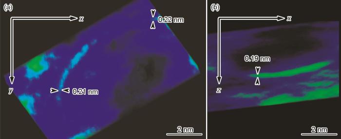
图6
表面Pt偏聚的3D探测尺寸
Fig.6
Detection of Pt segregation in x-y plane (a) and x-z plane (b) (The color represent the intensity of the EDS signal of the Pt element, which gradually becomes more intense from blue to green)
4 结论
(1) 利用原子分辨率的电子层析技术分析了纳米多孔金孔棱表面的原子结构,发现表面缺陷可以分为2类,一类是{111}平台上的扭折和台阶,一类是表面凹坑。相比于扭折和台阶,表面凹坑会引入更多的低配位位点。
(2) 将原子级电子层析技术与EDS成分三维重构相结合,分析了Pt元素在孔棱表面的偏聚,显示了Pt在孔棱表面低配位位点的偏聚会阻碍纳米多孔金属孔棱的粗化。原子分辨率的电子层析技术和三维化学成分分析为同时从原子尺度对材料的晶体学和化学成分进行分析提供了有效手段。
参考文献
Nanoporous gold: A new gold catalyst with tunable properties
[J].
Nanoporous gold (np-Au) represents a novel nanostructured bulk material with very interesting perspectives in heterogeneous catalysis. Its monolithic porous structure and the absence of a support or other stabilizing agents opens up unprecedented possibilities to tune structure and surface chemistry in order to adapt the material to specific catalytic applications. We investigated three of these tuning options in more detail: change of the porosity by annealing, increase of activity by the deposition of oxides and change of activity and selectivity by bimetallic effects. As an example for the latter case, the effect of Ag impurities will be discussed. The presence and concentration of Ag can be correlated to the availability of active oxygen. While for the oxidation of CO the activity of the catalyst can be significantly enhanced when increasing the content of Ag, we show for the oxidation of methanol that the selectivity is shifted from partial to total oxidation. In a second set of experiments, two different metal-oxides were deposited on np-Au, praseodymia and titania. In both cases, the surface chemistry changed significantly. The activity of the catalyst for oxidation of CO was increased by up to one order of magnitude after modification. Finally, we used adsorbate controlled coarsening to tune the structure of np-Au. In this way, even gradients in the pore- and ligament size could be induced, taking advantage of mass transport phenomena.
Unsupported nanoporous gold for heterogeneous catalysis
[J].
Nanoporous Au-Pt alloys as large strain electrochemical actuators
[J].
Charge-induced reversible strain in a metal
[J].Dimension changes on the order of 0.1% or above in response to an applied voltage have been reported for many types of materials, including ceramics, polymers, and carbon nanostructures, but not, so far, for metals. We show that reversible strain amplitudes comparable to those of commercial piezoceramics can be induced in metals by introducing a continuous network of nanometer-sized pores with a high surface area and by controlling the surface electronic charge density through an applied potential relative to an electrolyte impregnating the pores.
Nanoporous gold: A new material for catalytic and sensor applications
[J].Nanostructured materials are governed by their surface chemical properties. This is strikingly reflected by np-Au. This material can be generated by corrosion of bulk Ag-Au alloys. Based on a self-organisation process, a 3 dimensional sponge like gold structure evolves with ligaments in the range of only a few tens of nanometers. Due to its continuous porosity, the material can be penetrated by gases which then adsorb and interact with the surface. In this perspective we will review potential applications of np-Au resulting from this effect, namely heterogeneous gas phase catalysis, surface chemistry driven actuation, and adsorbate controlled stability of the nanostructure. We will summarize the current knowledge about the low temperature oxidation of CO as well as the highly selective oxidation of methanol. Furthermore, we will address the question how surface chemistry can influence the material properties itself. In particular, we will deal with (a) the actuation of np-Au by the reversible oxidation of its surface using ozone and (b) the adsorbate controlled coarsening of ligaments, using annealing experiments under ozone or inert gas atmosphere.
Fabrication and surface functionalization of nanoporous gold by electrochemical alloying/dealloying of Au-Zn in an ionic liquid, and the self‐assembly of L‐cysteine monolayers
[J].
Li Storage in 3D nanoporous Au‐supported nanocrystalline tin
[J].
Evolution of nanoporosity in dealloying
[J].
Characteristic features of alloy polarization curves
[J].
Defects evolution in nanoporous Au (Pt) during dealloying
[J].
Corrosion‐induced strengthening: Development of high‐strength nanoporous metals
[J].
Nanoporous metals by alloy corrosion: Formation and mechanical properties
[J].Nanoporous metals prepared by the corrosion of an alloy can take the form of monolithic, millimeter-sized bodies containing approximately 1015nanoscale ligaments per cubic millimeter. The ligament size can reach down to the very limits of stability of nanoscale objects. The processes by which nanoporous metals are formed have continued to be fascinating, even though their study in relation to surface treatment, metal refinement, and failure mechanisms can be traced back to ancient times. In fact, the prospect of using alloy corrosion as a means of making nanomaterials for fundamental studies and functional applications has led to a revived interest in the process. The quite distinct mechanical properties of nanoporous metals are one of the focus points of this interest, as relevant studies probe the deformation behavior of crystals at the lower end of the size scale. Furthermore, the coupling of bulk stress and strain to the forces acting along the surface of nanoporous metals provide unique opportunities for controlling the mechanical behavior through external variables such as the electrical or chemical potentials.
Fragmentation of nanowires driven by Rayleigh instability
[J].Rayleigh instability of copper nanowires has been experimentally demonstrated. After annealing 30–50-nm-diam wires at temperatures between 400 and 600°C, different stages of the fragmentation process are observed by scanning electron microscopy. At 400°C, the wires start to fragment, forming shorter sections at 500°C, and finally decaying into a chain of nanospheres at 600°C. Average diameter and spacing of the spheres are in agreement with theoretical predictions. The Rayleigh instability applied to nanowires provides a structuring technique producing long chains of nanospheres, which should find interesting applications, for instance, by guiding light below the diffraction limit via coherent coupling of surface-plasmon polaritons.
Ultrafine nanoporous gold by low-temperature dealloying and kinetics of nanopore formation
[J].
Sign-inverted surface stress-charge response in nanoporous gold
[J].
A theory of secondary alloying effects on corrosion and stress-corrosion cracking
[J].
Stabilized nanoporous metals by dealloying ternary alloy precursors
[J].
An atomistic description of dealloying: Porosity evolution, the critical potential, and rate-limiting behavior
[J].
Nanoporous metals fabricated through electrochemical dealloying of Ag-Au-Pt with systematic variation of Au: Pt ratio
[J].
Beneficial effects of adsorbate-induced surface segregation of Pt in nanoporous metals fabricated by dealloying of Ag-Au-Pt alloys
[J].
Methanol electro-oxidation on nanoporous metals formed by dealloying of Ag-Au-Pt alloys
[J].
Nanoscale mechanism of the stabilization of nanoporous gold by alloyed platinum
[J].Nanoporous gold (NPG) is usually made by electrochemical dealloying of Ag from binary AgAu alloys. The resulting nanoscale ligaments are not very stable, and tend to coarsen with time by surface self-diffusion, especially in electrolyte, which may lead to inferior electrocatalytic properties. Addition of a small amount of Pt to the precursor alloy is known to refine and stabilize the nanoporous product (NPG-Pt). However, the mechanisms by which Pt serves to refine the microstructure remain poorly understood. The present study aims to expand our knowledge of the role of Pt by examining NPG-Pt at atomic resolution with Atom Probe Tomography (APT), as well as by aberration-corrected Transmission Electron Microscopy. Atomic level observation of Pt enrichment on ligament surfaces sheds light on the underlying mechanisms that give rise to Pt's refining effect. Owing to improved Ag retention with higher Pt content, NPG-Pt (made by dealloying AgAuPt) was shown to have the highest surface area-to-volume ratio, compared to NPG-Pt (made by dealloying AgAuPt). Quantitative estimates reveal up to 5-fold enrichment of Pt at nanoligament surfaces, compared to the precursor content, in NPG-Pt. The interface between the dealloyed layer and the substrate was captured by APT, for the first time. The findings of this investigation add insight into the functionality of NPG-Pt and its prospective catalytic performance.
Low-coordination sites in oxygen-reduction electrocatalysis: Their roles and methods for removal
[J].Low-coordination sites, including edges, kinks, and defects, play an important role in oxygen-reduction electrocatalysis. Their role was studied experimentally and theoretically for various Pt surfaces. However, the roughness effect on similar-sized nanoparticles that could elucidate the role of low-coordination sites has attracted much less attention, with no studies on Pd nanoparticles. Here, using Br- adsorption/desorption, we introduce an effective approach to reduce surface roughness, yielding Pd nanoparticles with smoother surfaces and an increased number of (111)-oriented facets. The resulting nanoparticles have a slightly contracted structure and narrow size distribution. Pt monolayer catalysts that contain such nanoparticles as the cores showed a 1.5-fold enhancement in specific and Pt mass activities for the oxygen reduction reaction compared with untreated ones. Furthermore, a dramatic increase in durability was observed with bromide-treated Pd(3)Co cores. These results demonstrate a simple approach to preparing nanoparticles with smooth surfaces and confirm the adverse effect of low-coordination sites on the kinetics of the oxygen-reduction reaction.© 2011 American Chemical Society
In situ surface imaging: high temperature environmental SEM study of the surface changes during heat treatment of an Al-Si coated boron steel
[J].
Nanoscale terahertz STM imaging of a metal surface
[J].
Collective radical oligomerisation induced by an STM tip on a silicon surface
[J].Over the past decade, on-surface fabrication of organic nanostructures has been widely investigated for the development of molecular electronic components, catalysts, and new materials. Here, we introduce a new strategy to obtain alkyl oligomers in a controlled manner using on-surface radical oligomerisations that are triggered by electrons between the tip of a scanning tunnelling microscope and the Si(111)√3 ×√3 R30°-B surface. This electron transfer event only occurs when the bias voltage is below -4.5 V and allows access to reactive radical species under exceptionally mild conditions. This transfer can effectively 'switch on' a sequence leading to the formation of oligomers of defined size distribution thanks to the on-surface confinement of the reactive species. Our approach enables new ways to initiate and control radical oligomerisations with tunnelling electrons, leading to molecularly precise nanofabrication.
Atomic-scale insights into surface species of electrocatalysts in three dimensions
[J].
3D microstructure analysis of silicon-boron phosphide mixed nanocrystals
[J].The microstructure of boron (B) and phosphorus (P) codoped silicon (Si) nanocrystals (NCs), cubic boron phosphide (BP) NCs and their mixed NCs (BxSiyPz NCs) has been studied using atom probe tomography (APT), transmission electron microscopy (TEM), and Raman scattering spectroscopy. The BxSiyPz NCs inherit superior properties of B and P codoped Si NCs such as high dispersibility in aqueous media and near infrared (NIR) luminescence and those of cubic BP NCs such as high chemical stability. The microanalyses revealed that BxSiyPz NCs are composed of a crystalline core and an amorphous shell. The core possesses a lattice constant between that of Si (diamond-cubic) and BP (cubic). The amorphous shell is comprised of B, Si and P, though the composition is not uniform and there are local B-rich, Si-rich and P-rich domains connected contiguously. The amorphous shell is proposed to be responsible for their superior chemical properties such as high dispersibility in polar solvents and high resistance to acids, and the crystalline core is responsible for the stable NIR luminescence.
Multishell intermetallic onions by symmetrical configuration of ordered domains
[J].
Direct subangstrom measurement of surfaces of oxide particles
[J].
Reversible wurtzite-tetragonal reconstruction in ZnO (10
Transition of dislocation nucleation induced by local stress concentration in nanotwinned copper
[J].Lu, N.; Du, K.; Lu, L.; Ye, H. Q. Chinese Acad Sci, Inst Met Res, Shenyang Natl Lab Mat Sci, Shenyang 110016, Peoples R China.
Size-dependent grain-boundary structure with improved conductive and mechanical stabilities in sub-10-nm gold crystals
[J].
Free-standing monatomic thick two-dimensional gold
[J].Monolayer metal membranes have attracted research attention owing to their fascinating physical properties. Unlike layered materials with weak interlayer van der Waals bonding, metallic monolayer membranes are difficult to exfoliate due to strong metallic bonding between layers. Here, we fabricate free-standing monatomic-thick Au membranes and nanoribbons framed in bulk crystals using dealloying inside transmission electron microscope. The Au membranes are robust under high energy electron beam. Monatomic-thick nanoribbons with a minimal width of 0.6 nm are observed. First-principles calculations reveal that zigzag-edged nanoribbons are ferromagnetic with magnetic moments ranging 0.38-0.51 μB per unit-cell for a width less than 0.9 nm. In addition, a linear relationship between the bond length and the coordination number of atoms is directly investigated using atomic resolution images of monolayer and bilayer Au membranes. This work provides a pathway for direct fabrication of metal membranes and nanoribbons and to achieve novel physical properties.
Three-dimensional atomic imaging of colloidal core-shell nanocrystals
[J].Colloidal core-shell semiconductor nanocrystals form an important class of optoelectronic materials, in which the exciton wave functions can be tailored by the atomic configuration of the core, the interfacial layers, and the shell. Here, we provide a trustful 3D characterization at the atomic scale of a free-standing PbSe(core)-CdSe(shell) nanocrystal by combining electron microscopy and discrete tomography. Our results yield unique insights for understanding the process of cation exchange, which is widely employed in the synthesis of core-shell nanocrystals. The study that we present is generally applicable to the broad range of colloidal heteronanocrystals that currently emerge as a new class of materials with technological importance.
Electron tomography at 2.4-ångström resolution
[J].
Three-dimensional atomic structure of grain boundaries resolved by atomic-resolution electron tomography
[J].
3D atomic imaging of low-coordinated active sites in solid-state dealloyed hierarchical nanoporous gold
[J].
Surface energies of elemental crystals
[J].The surface energy is a fundamental property of the different facets of a crystal that is crucial to the understanding of various phenomena like surface segregation, roughening, catalytic activity, and the crystal’s equilibrium shape. Such surface phenomena are especially important at the nanoscale, where the large surface area to volume ratios lead to properties that are significantly different from the bulk. In this work, we present the largest database of calculated surface energies for elemental crystals to date. This database contains the surface energies of more than 100 polymorphs of about 70 elements, up to a maximum Miller index of two and three for non-cubic and cubic crystals, respectively. Well-known reconstruction schemes are also accounted for. The database is systematically improvable and has been rigorously validated against previous experimental and computational data where available. We will describe the methodology used in constructing the database, and how it can be accessed for further studies and design of materials.
Porous gold with a nested‐network architecture and ultrafine structure
[J].
