Sm2Co17磁体胞状组织边缘2:17R'相的透射电子显微镜表征
Identification of 2:17R' Cell Edge Phase in Sm2Co17-Type Permanent Magnets by Transmission Electron Microscopy
Sm2Co17磁体胞状组织边缘2:17R'相的透射电子显微镜表征 |
| 陈虹宇, 宋欣, 周相龙, 贾文涛, 袁涛, 马天宇 |
|
Identification of 2:17R' Cell Edge Phase in Sm2Co17-Type Permanent Magnets by Transmission Electron Microscopy |
| CHEN Hongyu, SONG Xin, ZHOU Xianglong, JIA Wentao, YUAN Tao, MA Tianyu |
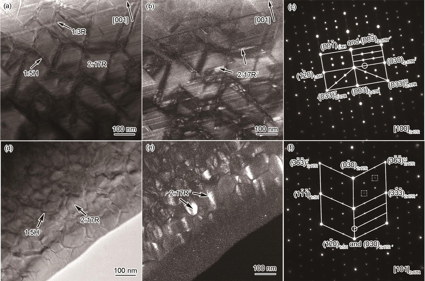
| 图1 沿Sm25Co50.2Fe16.2Cu5.6Zr3.0磁体[100]2:17R和[101]2:17R晶带轴的TEM明、暗场像及选区电子衍射(SAED)花样 |
| Fig.1 TEM bright field images (a, d), dark field images (b, e), selected area electron diffraction (SAED) patterns (c, f) for the Sm25Co50.2Fe16.2Cu5.6Zr3.0 magnet taken along [100]2:17R (a-c) and [101]2:17R (d-f) zone axes (The dark field images were taken using the (010)2:17R' or (020)2:17R' superlattice reflections circled by white in Figs.1c and f) |
|
|